Novel Photon-Counting Detector Concept for High-Resolution Radiographic Imaging
Project # 23-032 | Year 1 of 3
Stuart Miller
Los Alamos Operations (LAO)
This work was done by Mission Support and Test Services, LLC, under Contract No. DE-NA0003624 with the U.S. Department of Energy, the NNSA Office of Defense Programs, and supported by the Site-Directed Research and Development Program. DOE/NV/03624–1926.
Abstract
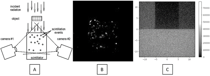
Neutron imaging has many important applications including those related to global security, such as Defense Nuclear Nonproliferation (DNN) and near-field detection, and many industrial applications in non-destructive testing (NDT) and radiography. Imaging neutrons is challenging due to their high penetrative power, which typically leads to low quality images with poor spatial resolution. Here we are developing a new imaging detector that will improve on both detection efficiency and spatial resolution. The innovative concept includes a thick transparent scintillator that is viewed by multiple cameras with lens coupling. The high-frame rate cameras are synchronized together with a short frame time to view individual scintillation events from multiple angles, which allows 3D spatial coordinates to be determined with great precision. The x, y, z values for thousands of frames and perhaps millions of events are compiled to form a high resolution 2D image. This innovative concept has the potential to impact not only global security, but a broad array of applications.
Background
There is a long-standing need within the response community to be able to determine the internal characteristics of an enclosed target. Traditional x‐ray techniques provide standoff capability to image the interior of the target and its contents. However, other key attributes can only be determined using methods that require physical contact with the enclosure or contents, which may not be allowable or possible. This project has explored the benefits that QCX may provide.
Technical Approach
The technical approach of this project was to first demonstrate the validity of this imaging concept. With this objective in mind, the following technical goals were identified:
- Identify and acquire suitable transparent scintillators for first imaging tests.
- Demonstrate 3D scintillator event imaging concept with gammas or X-rays.
- Work with Xcitex to evaluate software options and calibration grid design.
- Perform Groupe d’experts sur l’accès aux nouvelles technologies (GEANT) GEANT4 simulations with various scintillator configurations with gammas and neutrons of a range of energies.
- Develop image construction methods to convert the 3D event location data into a 2D radiographic image.
Three potential scintillators with high light yield were identified early in the project and samples were ordered for testing: thallium-doped cesium iodide, or (CsI:Tl), cerium-doped lutetium yttrium silicate (LYSO:Ce), and cerium-doped gadolinium aluminium gallium garnet (GAGG:Ce). Also, several framing cameras were identified including standard CMOS and electron-multiplying charge-coupled device (EMCCD) cameras. These cameras were borrowed for testing in the lab. It was quickly shown that EMCCD cameras were capable of imaging the scintillation events within standard CsI:Tl crystals with the lowest noise. Based on this, two EMCCD cameras were purchased.
Xcitex is a company that has developed software for 3D motion tracking with multi-camera images. A subcontract was placed with them to develop some modifications to their software specifically to locate and report the 3D scintillation event locations. Automating the process is essential to the program since it is not practical to go through thousands of frames to manually locate scintillation events.
GEANT4 simulations were performed with various configurations to demonstrate the theoretical performance of this concept with gammas and thermal and fast neutrons. While this imaging method eliminates the dark noise and read noise of the camera, the shot noise of the incident radiation is still present as well as scatter from the object and within the scintillator itself. These can be accounted for through these simulations with each species.
This new imaging technique also requires new methods to form the actual radiographic images. With normal radiographic imaging detectors, the images are formed by simply integrating for the required exposure time or adding multiple image frames together. Here, however, the x, y coordinates of the scintillation events are used to construct the image. A script was written in Python to project the x, y locations into an artificially generated TIF image based on the number of events at each x, y location. In addition, x, y coordinates can be chosen at different depths (z ranges) in the scintillator, allowing for energy-selective imaging.
Results and Technical Accomplishments
The results after the first year of this project are very promising. Perhaps the biggest accomplishment is that scintillation events can be resolved inside a bulk crystal scintillator. This was a milestone achievement because previous imaging of scintillation events was done with planar 2D scintillator screens, not focusing through a depth of transparent crystal.
The first year accomplishments were as follows:
- Identified suitable scintillators that work well for scintillation event imaging. The standard CsI:Tl scintillator works well for concept validation. This is a 25 mm thick monolithic detector.
- Evaluated several cameras, including CMOS, intensified CCD, and EMCCD technologies as well as a new quantitative CMOS (qCMOS) camera (a modified CMOS camera with ultra-low noise). The EMCCD cameras were the best match for this work and two cameras were purchased (a stretch goal for year 1).
- GEANT4 simulations were performed to demonstrate imaging techniques with gammas, thermal neutrons, and fast neutrons.
- Image construction methods were developed to make 2D radiographs from scintillation event location data provided by simulations.
- Software modifications (to provide x, y, z locations for scintillation events) for the Xcitex ProAnalyst 3D motion tracking software are underway.
- A complete two-camera EMCCD imaging system has been assembled in the lab to fully demonstrate the imaging technique.
Conclusions and Path Forward
From the work so far, we can conclude that this new imaging concept holds promise to become a high-performance radiographic imaging system. In the first year, we have shown that scintillation events can be resolved inside transparent scintillator crystals. A complete two-camera system has been purchased and set up in the lab. GEANT4 simulations have been performed and analysis is still underway. Image construction techniques, which are unique to this project, are currently in development.
The plan for completion of the project is to utilize the two-camera imaging system in the lab to demonstrate the full concept. Second year initial tests will include alpha and gamma event imaging of test objects with CsI:Tl and LYSO. The Xcitex software will be used to identify and provide location coordinates for the scintillation events. These data will be used to construct a high-resolution 2D radiograph using the image construction methods that have been developed.
With the completion of this initial real-world demonstration, the system can begin to be tested with thermal neutrons and fast neutrons. To accomplish this, suitable scintillators will be identified for each, and the imaging system can be taken to appropriate neutron sources for image acquisition and demonstration of the system. At the same time, we will work on optimizing and improving the imaging system. An optimized lens system will allow the smaller and dimmer scintillation events to be imaged. Also, adding anti-reflective coatings to the scintillators will help to enhance light collection efficiency to the cameras.
Working with Xcitex, we will also determine the best camera configuration in order to provide the most accurate scintillation event locations. In addition, we will estimate the expected improvements if we were to add a third camera to the system. A publication for this work is planned for the second year.