
Broadband X-Ray Imager for Spectroscopic Diagnostics
Project # 23-061 | Year 3 of 3
Radu Presuraa, Matthew S. Wallaceb, Showera H. Haqueb, Isiah D. Pohla, Padrick A. Beggsa, Alex L. Droemera
aSandia Operations (SO), bLivermore Operations (LO)
This work was done by Mission Support and Test Services, LLC, under Contract No. DE-NA0003624 with the U.S. Department of Energy, the NNSA Office of Defense Programs, and supported by the Site-Directed Research and Development Program. DOE/NV/03624–1883.
Abstract
This project aims to develop an instrument for two-dimensionally space-resolved diagnostics of mm- to cm-sized high energy density hot plasmas. The instrument is based on the property of convex crystals to diffract monochromatic x-rays stigmatically in the dispersion direction, over a broad spectral range. Adding a slit for imaging in the perpendicular direction produces a spectrum in which each line is an image of the x-ray source. Consequently, the spectroscopic diagnostics typically used to measure the plasma density and temperature can be applied point by point over the source projection. Building on previous work during the last year of the project, we evaluated the instrument performance using a centimeter-sized Henke x-ray source and demonstrated the ability to produce spectra consisting of source images in each spectral line. Through measurement and ray tracing modeling, we determined that the applicability of this technique can be extended to beam-target x-ray sources, as well as to radiographic applications.
Background
High energy density plasmas used for fusion and radiation source studies are commonly diagnosed using x‐ray spectroscopy (to measure the plasma temperature and density) and x‐ray imaging (to obtain the emissivity distribution). Combining these diagnostics provides 2D maps of plasma density and temperature useful to better understand the plasma dynamics and as verification for the results of radiation hydrodynamics codes. The uncertainties inherent to this technique prompted the development of several types of imaging spectrometers. Arrays of concave (spherical or toroidal) crystals in off‐Rowland circle geometries provide the highest spectral and spatial resolution. The spectral coverage is very limited, approximately one spectral line width per crystal. A different approach, called a Multi‐Monochromatic Imager, uses a densely packed pinhole array for imaging coupled with a flat Bragg reflector for spectral dispersion. This instrument provides comparatively lower spectral and spatial resolution over several spectral lines. It is also cheaper and easier to align and operate than the concave crystal arrays. Its applicability is restricted to mm‐sized x‐ray sources. However, certain applications require broader spectral coverage or diagnostics of larger x-ray sources. The most basic spectroscopic diagnostics use intensity ratios of pairs of spectral lines to determine the plasma density and temperature. These methods can be subject to large errors for non-equilibrium or anisotropic plasmas, which affect the populations of the upper transition levels; better results can be obtained by using more spectral lines, over a broad spectral range. Additionally, larger, centimeter-sized x-ray-plasmas are often inhomogeneous, with the density and temperature varying substantially from point to point. In this case, obtaining a single value integrated over the plasma volume for each plasma parameter is inadequate for comparing with the modeling codes; 2D density and temperature distributions are beneficial for plasma diagnostics.
Technical Approach
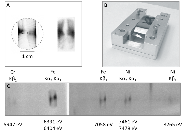
Our contribution is a different type of x‐ray polychromatic imager (XPI), based on a rarely appreciated property of convex Bragg reflectors: the spectral line profiles produced are sensitive to the source emissivity profile. In an optimized measurement setup, this instrument produces a stigmatic image of the x‐ray source in each spectral line, over a broad spectral range. While this XPI provides lower spectral and spatial resolution than the other methods, its spectral coverage is the widest, which is advantageous for spectroscopic diagnostics. Additionally, it is applicable to centimeter‐sized sources, such as z‐pinches, which distinguishes it from the previous solutions. Finally, it is cheaper and easier to set up. The central component of the XPI is a crystal bent as a segment of circular cylinder with its convex surface facing both the x‐ray source and the detector. This is the same geometry used in opacity measurements at Z and in the original configuration of the opacity spectrometer developed for the National Ignition Facility. In those applications, the instrument is optimized for spectral resolution. The XPI developed under this project can provide a complementary function, namely measuring the x-ray object’s 2D spatial distribution, although without enough spectral resolution to measure line profiles. To image in the direction perpendicular to the spectral dispersion plane (DP), the instrument uses a slit, as usual. Imaging in the DP can be optimized by reducing the distance from the x‐ray source to the crystal and the crystal curvature radius, and by increasing the distance between the crystal and the detector. The parameter values are determined by the x-ray source size and must be selected such that the image magnification is maximized, while avoiding the overlap of neighboring spectral lines. Emphasizing the imaging capability limits the spectroscopic functionality of the XPI, and any diagnostics based on line profiles are precluded. During the last year of the project, we assessed the concept using a laboratory x-ray source. We designed and built several crystal bending fixtures to work with several bending radii and to evaluate the effect of the bending method on the actual shape of the crystal and its imaging properties. The evaluation included two crystals, potassium acid phthalate (KAP 001) and silicon (Si 100), which have different elastic constants, crystalline structure, and lattice perfection degree. We used a centimeter-sized electron beam-target x-ray source, namely the Henke source housed at the NNSS’ Livermore Operations, with two different anodes. A silver alloy anode produced characteristic lines in the 1.8-3.5 keV range, and a stainless-steel anode emitted spectral lines between 6 and 8.3 keV. Both crystals were 100‑µm thick slabs, and the bending radii for KAP and Si were 4″ and 5″, respectively. The width of the slit used for imaging orthogonal to the dispersion direction was between 0.5 and 1 mm. The main result of the measurements was that the instrument can be easily set up to produce monochromatic source images in each spectral line over a broad spectral range. In the case of the Si 100 crystal, only the surface produced a spectrum, in the first allowed diffraction order (n = 4). For the KAP 001, with many highly reflective planes, the spectrum was complex and difficult to interpret even in the surface dispersion plane. In both cases, using 2D ray tracing enabled us to identify the spectral lines and the planes diffracting them. The measured spectra showed that it is not always possible to prevent spectral line overlap when optimizing the instrument for imaging.
Results and Technical Accomplishments
We demonstrated, with laboratory measurements, that the XPI has monochromatic imaging ability in each spectral line over a broad spectral range for a centimeter-sized x-ray source. The tests included two very different crystals, KAP and Si, and a total spectral range extending from 2 keV to 8 keV. Our 2D analytical calculations and 3D ray tracing modeling, developed in previous years, were useful in setting up the instrument and identifying both the spectral lines observed and the crystal planes responsible for their diffraction. The experimental data was successfully used to verify the 2D ray tracing software that we developed previously for instrument design and data analysis. The crystal bending fixture design proved to be effective and will continue to be used. Bent crystal diffraction calculations developed to select crystalline planes for this project are being used to support bent crystal calibrations for Sandia diagnostics. Results of this work were presented at four conferences and a manuscript is in preparation.
Conclusions and Path Forward
This project demonstrated that it is possible to produce a spectrum of an x-ray source in which each spectral line is an image of the source. Using this technique to measure the 2D distribution of density and temperature in high energy density hot plasmas can enable improved verifications of the plasma modeling codes. While Sandia appreciates the opportunity to use our crystal configuration, they asked to evaluate other crystal configurations for XPI applications. During this project, it became apparent that this technique can be applied to measure linear induction accelerator-driven x-ray source sizes, using fluorescent radiation from the electron beam target. A proposal for this application was submitted to the SDRD program. Recently, we realized that this technique can be adapted for radiographic measurements: in certain circumstances, a uniform, polychromatic x-ray source larger than the object investigated can be used to determine its composition and structure. In all applications mentioned, the source characteristics may change rapidly in time, and time integration may corrupt the measurements. The instrument developed can be equipped with a time-gated x-ray imager, such as an hCMOS framing camera, to add time resolution.
Publications
- Title: Multi-monochromatic Imaging a Henke X-ray Source with a Cylindrical Convex Crystal
Journal / Conference: 50th International Conference on Plasma Science, May 21-25, 2023, Santa Fe, NM
Year: 2023
Author(s): R. Presura, M. S. Wallace, S. H. Haque, J. M. Heinmiller, P. A. Beggs, I. D. Pohl - Title: Multi-monochromatic Imaging with Cylindrically Bent Convex Crystals
Journal / Conference: 24th High Temperature Plasma Diagnostics Conference, May 15-19, 2022, Rochester, NY
Year: 2022
Author(s): R. Presura, M. S. Wallace, S. H. Haque, J. M. Heinmiller, P. A. Beggs, I. D. Pohl, A. L. Droemer, P. Lake, M. Wu - Title: Calibrations of Quartz (10-11) in both Laue and Bragg diffractions between 8-45 keV Using Micro-focusing X-Ray Sources in Air
Journal / Conference: 24th High Temperature Plasma Diagnostics Conference, May 15-19, 2022, Rochester, NY
Year: 2022
Author(s): P. W. Lake, G. P. Loisel, R. Presura, T. J. Webb, K. J. Moy, M. Wu - Title: Broadband 2D Imaging Spectroscopic Diagnostic Techniques, poster UO6-4
Journal / Conference: 63rd Annual Meeting of the APS Division of Plasma Physics, November 8-12, 2021, Pittsburg, PA
Year: 2021
Author(s): S. Haque, R. Presura, M. S. Wallace, I. Pohl, J. Heinmiller - Title: Multi-monochromatic X-ray Imaging with a Cylindrical Convex Crystal for Spectroscopic Diagnostics
Journal / Conference: 65th Annual Meeting of the APS Division of Plasma Physics, October 30-November 3, Denver, CO
Year: 2023
Author(s): R. Presura, M. S. Wallace, S. H. Haque, I. D. Pohl, A. Droemer, J. M. Heinmiller, P. A. Beggs