
Non-Invasive Spot Size Diagnostic for Linear Induction Accelerators
Project # 23-058 | Year 1 of 1
Evan R. Scotta, Showera Haqueb, Robert Hibbarda, Stuart R. Millerc,
Aimee Neilsenb, Zachary C. Shawa, Michael E. Wellera, Trevor J. Burris-Moga, The DARHT Physics Teamd
aNevada National Security Sites (NNSS), bLivermore Operations (LAO), cLos Alamos Operations (LAO), dNational Lab: LANL
This work was done by Mission Support and Test Services, LLC, under Contract No. DE-NA0003624 with the U.S. Department of Energy, the NNSA Office of Defense Programs, and supported by the Site-Directed Research and Development Program. DOE/NV/03624–1882.
Abstract
Radiographic performance of a linear induction accelerator (LIA) can be largely described by two key parameters: the spot size on the converter target and the dose delivered to the object being radiographed. Currently, the spot size during radiographic measurements using LIAs is inferred from a series of invasive measurements that cannot be performed concurrently with the radiograph. To address this issue, a non-invasive beam spot size diagnostic has been developed for measuring upstream-propagating Bremsstrahlung x-rays from the converter target using an aperture, scintillator, and intensified charged coupled device (ICCD) camera. Testing of a variety of scintillator materials was performed with a 450 kV Bremsstrahlung source using a Complementary Metal Oxide Semiconductor (CMOS) camera. The diagnostic was demonstrated on Axis 2 of the Dual-Axis Radiographic Hydrodynamic Test facility (DARHT) where preliminary data were gathered during focused shots. Though an image is clearly visible, signal-to-noise must be improved to reduce overall uncertainty in the measurement. Additional x-ray shielding between the beam pipe and the scintillator is planned for future measurements.
Background
LIAs are commonly used to produce short, high-intensity bursts of x-rays for radiographic measurements. This is achieved by directing an intense relativistic electron beam into a high-Z material, thereby creating Bremsstrahlung x-rays. These x-rays are then used to image dense matter that would be opaque to shorter wavelengths.
The performance of an LIA can be quantified by the integrated spot size on the x-ray converter during one of these pulses and the x-ray dose generated by the electrons’ interactions with the converter. To determine the spot size on the converter, a series of invasive spot size measurements are taken before the radiographic measurement. However, due to the invasive nature of these measurements, the spot size during a radiographic shot of interest cannot be measured during the shot of interest, and experimental verification is therefore not possible using these methods.
Kolesnikov (2020) demonstrated a non-invasive beam measurement using upstream-propagating x-rays from a Bremsstrahlung converter by using a rolled edge, scintillator, and camera. A Site-Directed Research and Development Feasibility Study performed by Weller in 2022 confirmed the possibility of these measurements using the particle-in-cell code, Chicago. This project aims to demonstrate a scintillator-based diagnostic for non-invasive beam spot size measurements on an LIA as a follow-on to the 2022 Feasibility Study.
Technical Approach
To develop a diagnostic capable of measuring the beam spot size during a single pulse of a multi-pulse LIA, the general design is that of an aperture, scintillator, and gated camera. To measure a single pulse in a multi-pulse LIA, signal-to-noise levels must be sufficiently high during a single pulse to resolve the image after the aperture. However, image resolution must be sufficiently high to resolve a spot on the order of millimeters. Because both the resolution and signal level are directly related to the scintillator, samples of multiple scintillators were tested using a 450 kV Bremsstrahlung source at Los Alamos National Laboratory’s TA‑08. Of these scintillators, LSO:Ce powder screens have the highest resolution and among the highest response, and were chosen for use in preliminary measurements at DARHT Axis 2.
To develop and demonstrate the source spot reconstruction technique used by Bachmann in 2016, an analysis of the 450 kV source penumbral imaging measurements was performed for each scintillator sample.
Though a mechanical diagnostic concept was developed, preliminary measurements on DARHT Axis 2 used a temporary setup including an imaging fiber bundle in lieu of optical coupling. To create space on the DARHT Axis 2 variable field-of-view, shielding between the scintillator and the beam pipe was removed. This shielding, however, is normally used to reduce background noise during upstream measurements performed at DARHT Axis 2 using an image plate for an image integrated over multiple pulses in a pulse train and should be used for future measurements to reduce background noise.
A PI-MAX® 4 Electron-Multiplying Charge Coupled Device (EMCCD) camera located nearby to the DARHT Axis 2 variable field-of-view was shielded using tungsten bricks and coupled to the imaging fiber bundle using a C-mount lens relay. A mirror after the scintillator plate was used to direct light upward and into the fiber bundle to prevent the direct illumination of the bundle with x-rays. With the current experimental setup, there is a limitation on the magnification achievable due to the fixed position of the aperture and physical constraints on the scintillator location. However, there is interest at DARHT to be able to adjust the magnification of these diagnostic measurements. To accommodate this, a re-entrant pipe was designed for DARHT Axis 2 that allows an aperture to be located closer to the target location, thereby changing the magnification of the diagnostic.
Results and Technical Accomplishments
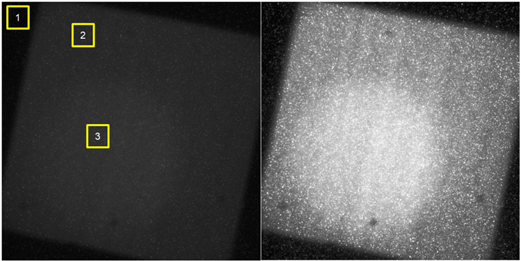
Measurements were taken during focused shots on DARHT Axis 2. The captured image is plagued by high background noise; without contrast-enhancing the raw image, very little is visible. However, this image proves that use of a gated camera and scintillator can give a non-invasive beam spot measurement.
With the current experimental setup, there is a limitation on the magnification achievable due to the fixed position of the aperture and physical constraints on the scintillator location. However, there is interest at DARHT to be able to adjust the magnification of these diagnostic measurements. To accommodate this, a re-entrant pipe was designed for DARHT Axis 2 that allows an aperture to be located closer to the target location, thereby changing the magnification of the diagnostic.
Conclusions and Path Forward
Preliminary measurements at DARHT Axis 2 have shown a scintillator-based diagnostic can be used to measure upstream-propagating x-rays resulting from the electron beam interaction with the converter target. Though signal-to-noise in the preliminary measurements is low, improvements to the diagnostic can likely address these issues to provide accurate measurements of the beam spot during radiographic measurements. The primary improvement is the assimilation of additional shielding between the beam pipe and the scintillator.
Future measurements may be performed again with an imaging fiber bundle or with optical coupling. An additional measurement without the aperture would allow for the characterization of features within the scintillator that could be removed from the final image. If an imaging fiber is used, at least one measurement without the scintillator present should be taken to ensure no scintillation in the fiber confounds the measurement. If light levels and field-of-view remain large issues during these measurements, the system can use optical coupling in lieu of the imaging fiber to reduce losses in the fiber bundle if the final light pipe can remain sufficiently dark. Faster lenses can also be incorporated in an optically coupled system to further increase overall signal levels.
This system can also be converted to a multi-pulse detection system using a gated camera capable of capturing multiple images within approximately 1 microsecond using a gated dual frame camera. By including a pellicle mirror between the scintillator and the aperture, a second camera may allow measurements of four individual pulses without appreciably lowering signal-to-noise. Additionally, because this system uses a scintillator to convert x-rays to visible light, there is potential for 1D time-resolved measurements of the beam spot using a rolled edge, cylindrical lens, and streak camera. These results would be convolved with the scintillator decay, however, and additional characterization of the scintillator may be necessary.
Back to Accelerator Beam Science and Target Interactions Index